


 Unmatched Expertise
Our team of highly skilled cardiologists and surgeons brings together decades of experience and specialized training to provide you with the most advanced cardiac care.
 Patient-Centric Approach
We prioritize your well-being, crafting personalized treatment plans and ensuring a compassionate, supportive environment throughout your journey to recovery.
 World-Class Infrastructure
We are equipped with state-of-the-art technology and advanced facilities, enabling us to provide the most precise diagnostics and effective treatments available.
SPECIALISTS
Our Expert Team
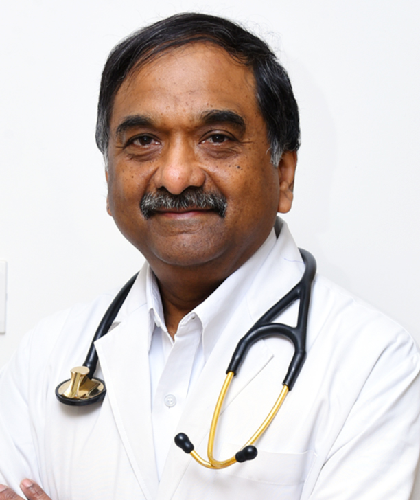
Dr. Thomas Alexander
Cardiologist
Dr. R. Sureshkumar
CardiologistDr. Minoti Subhash Kale
Paediatric CardiologistDr. M.Lawrance Jesuraj
Cardiologist, Clinical Cardiac ElectrophysiologistDr. M. Mohan
Interventional CardiologistDr. D.M.T. Saravanan
CardiologyDr. Balakumaran J
Consultant Interventional cardiologist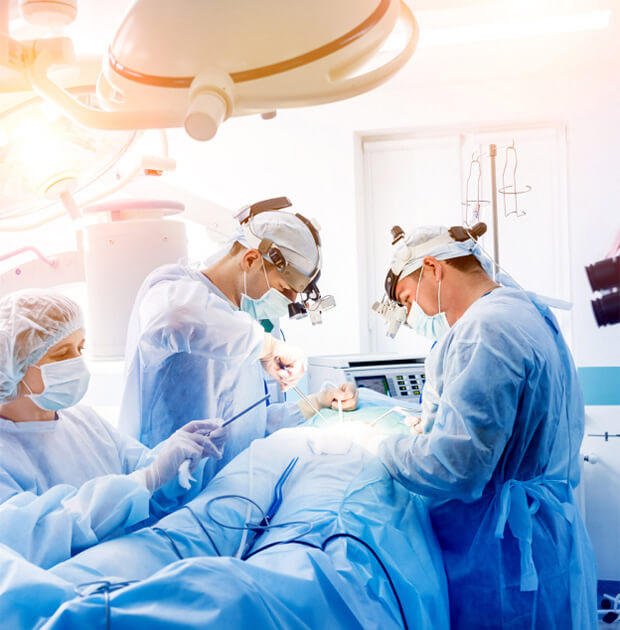
Lorem ipsum dolor sit amet, consectetur adipiscing elit. Ut elit tellus, luctus nec ullamcorper mattis, pulvinar dapibus leo.

Lorem ipsum dolor sit amet, consectetur adipiscing elit. Ut elit tellus, luctus nec ullamcorper mattis, pulvinar dapibus leo.

Lorem ipsum dolor sit amet, consectetur adipiscing elit. Ut elit tellus, luctus nec ullamcorper mattis, pulvinar dapibus leo.
